Against the background of painful processes in the lungs and other organs, it may occur. This pathology is not uncommon in medical practice, sometimes the symptoms of pleurisy come to the fore, masking the underlying disease.
Between the chest and lungs is a narrow space lined with a thin serous membrane - the pleura. The cavity contains about 2 ml of thick fluid, which ensures smooth movement of the lungs. This moisture is constantly filtered out of the blood vessels of the pleura. Fluid is absorbed through the lymphatic system and excreted to the lymph nodes.
Inflammatory processes are accompanied by the accumulation of effusion in the pleural cavity - an exudate rich in protein. The lymphatic vessels of the pleural sheets have time to remove a small amount of inflammatory fluid from the cavity, but fibrin clots settle on the surface of the serous membrane.
Fibrinous pleurisy develops, also called dry. With the accumulation of a significant amount of exudative fluid, the disease passes into an exudative form. When pathogens enter the pleural cavity, the inflammatory exudate becomes purulent. In addition to bacteria, the development of pleurisy can cause:
- fungal microorganisms;
- viruses;
- rickettsia;
- helminths;
- protozoa.
The infection reaches the pleural cavity through the circulatory and lymphatic system, or penetrates directly from the focus of the disease in the lungs. Infection can occur with an open chest injury, surgical intervention. The most common cause of infectious pleurisy is pneumonia.
Pleurisy, not associated with an infection, can provoke a lot of reasons, including:
- tumors (most often mesothelioma);
- vascular diseases;
- myocardial infarction;
- pancreatitis;
- kidney failure;
- radiation therapy;
- chest trauma.
Dry pleurisy
The main symptoms that the patient complains of with fibrinous pleurisy are fever and pain in the lesion of the pleura. The pain tends to increase during deep breaths, coughing, movement. In this case, a person puts his hand to a sore spot, as if protecting him.
Breathing becomes frequent, at times there is pain in the joints, muscles. Body temperature rarely reaches high values, accompanied by chills and excessive sweating.
When listening, the decisive diagnostic sign is the characteristic friction noise, which is produced by rough pleural sheets with layers of fibrin.  At the end of inhalation and the beginning of exhalation, a grinding sound is clearly defined, similar to the rustling of paper. At the site of the lesion, weakened, muffled breathing in the lungs is heard.
In a laboratory blood test, symptoms of inflammation are revealed: an increase in the number of leukocytes, an acceleration of the erythrocyte sedimentation rate, a shift in the white blood formula to the left. An X-ray examination shows a decrease in lung mobility, and an ultrasound shows deposits of fibrin flakes.
exudative form
In the initial stages of exudative pleurisy, a dry cough and soreness in the affected area of the pleura can be observed. As fluid accumulates in the pleural cavity, the pain disappears, but a feeling of fullness or heaviness in the chest appears. Body temperature often reaches 40˚, symptoms of intoxication are expressed:
- weakness;
- increased sweating;
- dizziness.
Inflammatory effusion displaces the organs of the chest cavity and disrupts the activity of the cardiovascular system. At the same time, the heart rate increases, blood pressure drops. Due to the infringement of lung function, shortness of breath develops, the patient's skin becomes cyanotic.
On examination, it is possible to detect an increase in the volume of the chest on the side of the lesion. Intercostal spaces over the area of inflammation are smoothed out, sometimes even protrude. The patient with pleurisy lies mainly on the affected side, as this relieves the pain a little. When tapping the place of accumulation of inflammatory exudate, a dull sound is heard. The fluid absorbs sounds, so breathing sounds in the lungs do not sound over the affected area.
are heard.
Purulent form
It can be quite difficult to recognize purulent inflammation of the pleural sheets. At the onset of the disease, symptoms of the underlying disease, such as pneumonia or acute pancreatitis, predominate. Increasing symptoms of intoxication indicate the attachment of infection to the inflammatory process: 

- weakness;
- lack of appetite;
- emaciation;
- trembling of the limbs;
- frequent pulse;
- exhausting fever.
Body temperature during the day can fluctuate significantly, falling sharply from higher to normal numbers. Temperature stabilization at the level of 37-37.5˚ is considered to be an extremely unfavorable sign. This indicates the depletion of the body's defenses. A cough develops with sputum production. The functions of the lungs, liver, kidneys and circulatory system are sharply disturbed. The skin of patients, especially on the face, becomes edematous.
The disease can be complicated by a breakthrough of the purulent contents of the pleural cavity into the bronchi. This is accompanied by the appearance of a large amount of sputum with an unpleasant odor. Much less often, a purulent effusion impregnates the soft tissues of the chest, subcutaneous tissue, thins the skin and breaks out.
In the blood test, there is a sharp increase in the number of leukocytes, a shift of the leukocyte formula to the left, neutrophils are affected by toxic granularity. Biochemical studies register an increase in the level of C-reactive protein, haptoglobin, sialic acids and other markers of acute inflammation.
Diagnostic studies
To confirm the diagnosis with exudative pleurisy, apply:
- radiograph;
- pleural puncture.
The initial stage of purulent pleurisy is best detected by x-ray examination of the chest in direct and lateral projections. With the accumulation of a large amount of inflammatory effusion, the radiograph is carried out in the position of the patient lying down. The fluid is evenly distributed along the chest and becomes visible in the picture.
The most difficult thing is to identify changes if the inflammation is accompanied by an adhesive process and the formation of a closed cavity with pus. It is possible to accurately establish the pathological accumulation of pleural fluid and determine the place for puncture using ultrasound.
Pleural puncture for exudative pleurisy is performed under local anesthesia. In the patient's sitting position, a puncture of the intercostal space is made with a long and thick needle. The liquid obtained during the puncture is subjected to: 
- microscopy;
- bacteriological analysis;
- cytological examination (to exclude a tumor).
Medical appointments
If any form of pleurisy is detected, treatment of the underlying pathology is necessary. With fibrinous pleurisy, treatment is supplemented with cough medicines (codeine, dionine) and painkillers. Symptoms of inflammation are relieved by Voltaren, Indomethacin or Acetylsalicylic acid.
At the first signs of fibrinous pleurisy, alcohol-containing compresses are effective. The prognosis for dry pleurisy is favorable, adequate treatment after 10-15 days leads to recovery, only sometimes there are relapses of the disease.
If the cause of exudative pleurisy cannot be established, then treatment with antibacterial agents is prescribed, as with pneumonia. When selecting drugs, only those that have not been used in the treatment of the patient before should be used.
Antibiotics from the penicillin group can be combined with cephalosporins, aminoglycosides or metronidazole to enhance the action. The drugs are best administered intravenously by drip.
Intravenous administration of hemodez, glucose, Ringer's solution with exudative pleurisy helps to reduce intoxication and correct protein metabolism. To enhance the reactivity of the body, treatment is supplemented with immunomodulating agents that activate phagocytosis and cause the production of interferon. Apply:
- Levamisole;
- T-activin;
- Sodium nucleinate;
- Timalin.
In the stage of resorption of the inflammatory effusion, paraffin therapy and chest massage are used. With the help of electrophoresis calcium chloride, heparin is introduced.
Treatment of pleurisy with drainage is carried out if:
- displacement of the heart with inflammatory fluid;
- symptoms of collapsed lungs (low blood pressure, severe shortness of breath);
- large amount of exudate.
A puncture is made and a tube with a valve is inserted along the needle. To avoid complications, no more than 1-1.5 liters of fluid is pumped out of the cavity at a time. Through the drainage, antibiotics are injected directly into the site of infection. If the treatment does not bring results, the affected areas of the pleural sheets and scar layers are surgically removed.
Each patient who has had pleurisy should be regularly examined by a general practitioner or pulmonologist within two years after the illness. Well restores the health of the respiratory system treatment in specialized sanatoriums.
It's cold time. At the same time, the number of patients with lung diseases is also increasing. Untreated bronchitis or pneumonia can often cause such an ailment as pleurisy, the symptoms and treatment of which everyone should know. After all, neglected forms of the disease sometimes lead to tuberculosis and oncology.
What is pleurisy?
The pleura is the membrane that covers the lungs. As a result of infections, adhesions or fluid may accumulate on its surface. This inflammatory process is called pleurisy.
Pleurisy occurs against the background of lung diseases as a complication. It can affect one or both parts of the respiratory organ. The disease occurs in an acute, chronic or mild form and is more common in children who have had pneumonia, as well as in tuberculosis patients. There are cases when the symptoms of the disease actually hide the pathology of the lungs or chest wall.
Treatment of pleurisy is always carried out permanently under the supervision of doctors. In severe cases, surgical intervention is indicated.
Causes and symptoms of the disease
The main cause of pleurisy is infection. The body produces antibodies that, interacting with microbes, damage the lining of the lungs, and this leads to the accumulation of fluid in its cavity and the formation of adhesions.
Microbes and toxins enter the pleura from foci of pneumonia, bronchitis, abscesses. Often pleurisy appears in patients with syphilis, brucellosis, tularemia, typhoid fever, and so on. Prerequisites may be the presence of bacteria such as fungi, mycoplasmas, blastomycosis.
Cause illness and non-infectious inflammatory processes in the body:
- Lung cancer, tumors of the breast, ovaries.
- Arthritis, rheumatism, scleroderma.
- Diseases of the heart and blood vessels (heart attack).
At risk are patients who:
- They got over the stress.
- Cooled down.
- Constantly overtired at work.
- They lead an unhealthy lifestyle, including malnutrition.
- Suffer from allergic reactions to chemicals.
The main signs of pleurisy:
- Pain that is especially noticeable when coughing.
- Hiccups, metabolism, night sweats.
- Crackles in the lungs, which are easily audible.
- High body temperature, fever, chills.
- Fatigue and feeling unwell.
- Dry and very prolonged cough.
- Shortness of breath, heaviness in the chest.
- Hemoptysis (sometimes).
Treatment of pleurisy of the lungs is performed only permanently. In order to avoid complications, including tumor formations, with similar symptoms, you should urgently go to the hospital. After a thorough diagnosis, the specialist will prescribe therapy. Patients who have had pleurisy should be seen by a doctor two to three years after full recovery.
Types of pleurisy
Depending on the course and nature of the disease, exudative pleurisy (accumulation of fluid in the pleural cavity) and fibrinous (formation of cysts and adhesions) are distinguished.
Exudative, in turn, is divided into:
- Serous - accumulation of serous fluid.
- Purulent - accumulation of pus.
- Putrid - accumulation of fluid with an unpleasant odor. This form is provoked by putrefactive microbes (gangrene of the lungs).
- Chylous - accumulation of lymph, which occurs due to squeezing of the lymphatic flow by the tumor.
The accumulated fluid can move (we are talking about diffuse pleurisy), or it can affect only one part of the pleura - encysted pleurisy.

In a separate group, cancerous pleurisy is distinguished - carcinomatous (damage to the membranes of the lung by cancer cells) and metastatic (metastases of the lung, pleura, breast and other organs).
Each of the forms of pleurisy is dangerous in its own way and requires immediate treatment. Ignoring the symptoms of the disease only leads to complications, the worst of which are malignant tumors.
Treatment of pleurisy occurs stationary with the help of complex therapy (antibiotics, vitamins, physical procedures). The duration of the disease can sometimes reach 4-6 months (chronic pleurisy). And it can take from ten to fourteen days (fibrinous pleurisy).
An accurate diagnosis can only be made by a doctor. Self-medication can only aggravate the patient's condition.
Exudative form of pleurisy
The disease is characterized by the accumulation of fluid (exudate) in the pleural cavity. Symptoms of exudative pleurisy appear slowly. However, pain is not a leading symptom of the disease. The patient mainly complains of feeling unwell, fatigue, headache, heaviness in the chest and shortness of breath.
The main cause of the disease in adults is tuberculosis and pneumonia. It is mainly possible to diagnose exudative pleurisy with the help of an x-ray, where fluid accumulations will be clearly visible. Treatment of this form of the disease includes antibiotic therapy and necessarily a diet that is aimed at limiting fluid intake.

When the amount of exudate is large enough, surgical intervention can be prescribed - a puncture, during which fluid is "sucked" from the lungs. The biomaterial must be examined in the laboratory, its nature is clarified and a final diagnosis is established.
Encapsulated pleurisy
One of the types of pleurisy, in which fluid accumulates in only one cavity of the pleura. Most often this occurs in the lower compartments of the lungs.
The patient complains of respiratory failure, high blood pressure, headaches, fever. When pressing on the chest, the patient experiences discomfort.

Complications of encysted pleurisy - bronchial fistula and empyema. A fistula is the formation of a pathological canal between the pleura and the organs of the chest. Empyema - accumulation of purulent fluid (purulent pleurisy).
The encysted form of pleurisy almost always develops with tuberculosis. Sometimes the patient is not even aware of its presence. Treatment of the disease depends on the degree of its neglect.
Fibrinous (dry) pleurisy
Fibrinous pleurisy, unlike exudative, is characterized not by the accumulation of fluid, but by the formation of adhesions on the surface of the pleura.
The main symptom is pain in the chest, fever, general weakness and exhaustion of the body. There is also a dry prolonged cough, rapid, shallow breathing.

When listening in the lungs, noises are heard that occur as a result of pleural friction.
Depending on the place of formation of cysts, there are:
- Diaphragmatic pleurisy (hiccups, painful swallowing, giving pain to the shoulder or abdominal wall).
- Apical - the apex of the lungs is covered with cysts. More often, this phenomenon accompanies tuberculosis (pain radiates to the shoulder, arm, shoulder blade).
- Paramediastinal - the anterolateral areas of the pericardium are affected (pain in the heart).
- Parietal (chest pain, aggravated by coughing). This is the most common form of dry pleurisy.
The main cause of the disease is exacerbation of forms of tuberculosis, pneumonia, less often bronchitis, typhoid fever, brucellosis.
Treatment of pleurisy (dry) is carried out quickly, within one and a half to two weeks after the onset of the disease.
Diagnosis and treatment
A residual diagnosis of "pleurisy" can be made after a doctor's examination and passing all the necessary tests.
Basic diagnostic methods:
- X-ray. Lesions will always be visible on x-rays. Therefore, for the purpose of prevention, it is recommended to undergo a medical examination and do fluorography of the lungs at least once a year.
- Puncture of the pleura - obtaining exudative fluid from the pleural cavity.
- Cytological examination of the fluid.
- Analysis of expectorant secretions (sputum).
- Lung ultrasound. It is necessary to assess the patient's condition and determine the tactics of treatment.
Treatment of pleurisy of the lungs occurs in two ways - with the help of chemotherapy and surgery.

Medicines are prescribed in a complex. In addition to antibiotics, the doctor prescribes antipyretics, painkillers and expectorants. Mandatory are immunostimulants and vitamins. It is very important to follow a diet, especially with exudative pleurisy. Physiotherapy, oxygen cocktails, massages can also be prescribed. Therapy is carried out only permanently under the supervision of a specialist doctor.
If the fluid accumulates very quickly and in large quantities, a mini-operation is prescribed, which is performed through a puncture. A pleurectomy (removal of the parietal pleura) can also be performed.
Folk ways to deal with the disease
Treatment of pleurisy can also be done with folk methods. However, they are more likely to be used as additional, but not as independent ones.
For oral administration:
- Mix one to one honey and onion juice. Take one tablespoon twice daily before meals.
- Honey with black radish juice (1:1). Drink one tablespoon three times a day.
- The root of the step (4 spoons) pour 500 ml of alcohol or vodka. Infuse for ten days in a warm place, then strain and drink one teaspoon three times a day, drinking (this is important!) Milk!
Compresses:
- The affected side should be rubbed with warm olive oil. After that, you need to wrap up well and lie down.
- Compress of mustard and honey. Take 30 g of mustard seeds, add a teaspoon of honey and pour water (2 cups). In the resulting solution, moisten a towel or gauze and apply a compress for twenty to thirty minutes. After the procedure, you need to dress warmly and relax.

Favorably on recovery affect massages and hiking in the fresh air. These activities are recommended to be carried out at the stage of recovery, but not during the acute course of the disease.
Complications of pleurisy
Pleurisy to a greater extent itself is a complication of many diseases associated with the respiratory tract. However, after it, problems can also arise that have no solution. This is respiratory and heart failure, which the patient receives as a result of thickening of the pleural membrane, or its fusion with other organs (with the heart, for example). The patient will experience such inconvenience for the rest of his life. In severe cases, surgery may be performed.
Of course, it is better not to bring your body to this stage of the disease. To do this, you need to seek help from a doctor in a timely manner, as well as undergo a scheduled examination annually.
Conclusion
Respiratory diseases are the most common in our time, especially among children. Therefore, people who often encounter various lung ailments must know what pleurisy is, the symptoms and treatment of the disease. Timely diagnosis will help to avoid further complications.
Pleurisy is an inflammation of the pleural sheets, accompanied by sweating into the pleural cavity of exudate of one nature or another. Sometimes the same term refers to non-inflammatory processes in the pleura, accompanied by the accumulation of pathological fluid in it (carcinomatous pleurisy, chilesian pleurisy), as well as irreversible changes in the pleura, which are the outcome of completed inflammation (adhesive pleurisy, ossifying pleurisy, etc.) . Usually, pleurisy is not an independent diseasebut is a pathological condition, complicatingflow of certain processes in the lungs and, much less frequently, in the chest wall, mediastinum, diaphragm and subdiaphragmatic space, or a manifestation of general (systemic) diseases, including those that occur without a distinct lesion of tissues in contact with the pleura. Despite the secondary nature of almost all inflammatory and reactive processes in the pleura, the latter are distinguished by the originality of clinical manifestations, often determine the course and severity of the underlying disease, and in some cases require special therapeutic measures. This justifies a separate consideration of pleurisy among other diseases of the respiratory system.
There are no reliable statistics regarding the frequency of pleurisy and mortality from them, since in most cases pleurisy is recorded under the headings of the main diseases that they complicate, and are often masked by other manifestations of the latter and are not recognized at all. Pleural adhesions, which are evidence of a past inflammatory process in the pleura, were found during autopsies in 48% of those who died from accidents, and in 80.5% of those who died from various diseases.
What causes pleurisy
All pleurisy can be divided into 2 large groups: a) infectious, i.e. associated with the invasion of the pleura by infectious pathogens, and b) non-infectious, or aseptic, in which the inflammatory process in the pleura occurs without the direct participation of pathogenic microorganisms.
From infectious etiological factors the most important are the causative agents of acute pneumonia and acute pulmonary suppuration, which are often complicated by an infectious process in the pleura (pneumococcus, staphylococcus, gram-negative rods, etc.). Tuberculosis microbacteria are also an important cause of pleurisy, and if until the middle of the current century tuberculosis was the most common cause of exudative pleurisy, in recent decades this has been noted in 20% of patients. Pleurisy of fungal etiology is known (with coccidioidomycosis, blastomycosis and other rare fungal infections).
Aseptic pleurisy may be of a very different nature. So, aseptic inflammation in the pleura can be the result of hemorrhage into the pleural cavity due to trauma or surgery (traumatic pleurisy), with penetration into the pleural cavity of invasive pancreatic enzymes as a result of acute pancreatitis (enzymatic pleurisy). Very often there are pleurisy associated with dissemination in the pleura, primary or secondary malignant tumors (carcinomatous pleurisy due to cancer metastases or pleural mesothelioma). Currently, it is pleural carcinomatosis that is the cause of up to 40 and even more percent of all exudative pleurisy.
Relatively often, aseptic pleurisy occurs as a result of a pulmonary infarction. Known aseptic pleurisy in systemic diseases of the connective tissue (rheumatism, "big" collagenosis), as well as in leukemia, Hodgkin's disease, hemorrhagic diathesis (Werlhof's disease), in some diseases of the kidneys and liver. It should, however, be noted that not all of the listed cases have an inflammatory nature. Pleural changes seem indisputable.
Pathogenesis (what happens?) during Pleurisy
Ways of penetration of microorganisms into the pleural cavity. lost at infectious pleurisy may be different. Apparently, it is important direct inpleura infection from subpleurally located pulmonary foci. Tuberculous, pleurisy is characterized by seeding of the pleural cavity from the radical lymph nodes, subpleural foci, or as a result of a breakthrough of caverns with the formation of pyopneumothorax. Retrograde flow of tissue fluid from the depths to the surface of the lung, in all likelihood, can cause lymphogenous infection pleural cavity. Microbial contamination of the pleura by hematogenous route is of lesser importance and occurs mainly indirectly, through the formation of foci of hematogenous infection in the subpleural layer of the lung. Finally, in surgical practice, the main role is played by direct infection pleura from the external environment during injuries and surgical interventions, as well as as a result of opening intrapulmonary purulent foci during surgery.
The development of the infectious process in the pleural cavity is determined by the following circumstances: firstly, the very fact of its infection and the characteristics of the infection; secondly, the peculiarities of the local and general reactivity of the patient; thirdly, local conditions created in the pleural cavity during its infection. In some cases, for example, with nonspecific purulent pleurisy, the main role is played by the ingress of pathogens (pus) into the pleural cavity. With tuberculous pleurisy, sensitization under the influence of the previous course of a specific process is very important, as a result of which the ingestion of an insignificant amount of mycobacteria leads to a hyperergic reaction with a rapid accumulation of exudate, in which pathogenic microbes can only be detected with great difficulty. Such pleurisy is considered infectious-allergic.
An important role in the development of pleurisy, especially purulent, is also played by local conditions in the pleural cavity, in particular the accumulation of air or blood in it, which is an excellent medium for the development of pyogenic microflora.
Pathogenesis non-infectious pleurisy studied to a lesser extent. Aseptic traumatic pleurisy is associated with the reaction of the pleura to the outflow of blood, which usually does not coagulate with small hemothoraxes, gradually dilutes with accumulating exudate and subsequently resolves, leaving relatively small adhesions. With a large hemothorax and severe trauma to the chest wall and lung, the blood in the pleural cavity coagulates (clotted hemothorax). In the future, if suppuration does not occur, a massive clot undergoes organization by connective tissue, resulting in the formation of thick ligaments that limit lung function.
The so-called sympathetic or sympathetic pleurisy is associated with exposure to the pleura of toxic products from nearby infectious foci, as well as with lymphogenous invasion of pancreatic enzymes in pancreatitis. The aseptic flowing so-called parapyeumonic pleurisy can also be attributed to the same type. It should be noted that the traditional division of pleurisy associated with pneumonia into para- and metapneumonic ones is not entirely correct, since with metapneumonic pleurisy it is usually not an independent process that occurs after the resolution of pneumonia, but about secondary infection and suppuration that appeared in the midst of pneumonia of aseptic reactive (parapneumonic) effusion, which was not recognized in a timely manner.
The effusion in carcinomatous pleurisy is associated, on the one hand, with the impact on the pleura of the products of pathological tumor metabolism, and on the other hand, with a violation of the circulation of the lymph as a result of the blockade of the ways of its outflow (regional lymph nodes, the so-called "hatches" of the parietal pleura) neoplasm elements.
The pathogenesis of pleural exudation in collagen diseases is obviously associated with systemic vascular damage and changes in the general reactivity of patients.
The mechanism of formation and evolution of pleural exudate seems to be quite complicated. The physiological flow of tissue fluid through the pleural cavity in the direction from the surface of the lung to the chest wall causes the fact that with the remaining outflow and moderate effusion, the liquid part of the latter can be resorbed, and only a dense layer of fibrin precipitated from the exudate remains on the surface of the pleura, resulting in the formation fibrinous or dry pleurisy. If the rate of exudation begins to exceed the capacity of the outflow, which can be blocked as a result of inflammation, liquid exudate accumulates in the pleural cavity, squeezing the lung, and the pleurisy becomes serous-fibrinous or, if fibrin does not fall out, serous. With the reverse development of the process, when the rate of resorption begins to prevail over the rate of exudation, the liquid part of the effusion is absorbed, and the fibrinous deposits are organized by the connective tissue with the formation of moorings, the massiveness of which determines the subsequent violation of respiratory functions, and with partial or complete obliteration of the pleural cavity.
At exudate infection pyogenic microflora, the latter acquires serous-purulent, and then purulent character, and formed pleural empyema. Under no circumstances can purulent exudate be resorbed, and its elimination can occur only as a result of a breakthrough to the outside (with purulent fusion of chest wall tissues), through the bronchial tree, or as a result of therapeutic effects (puncture, drainage of the pleural cavity).
Along with fibrinous, serous-fibrinous and purulent effusions, there are other types of exudates in pleurisy. So, with pleural carcinomatosis, pulmonary infarction, with pancreatitis, occasionally with tuberculosis and in some other conditions, hemorrhagic exudate. In allergic processes, eosinophils may predominate in the effusion. (eosinophilic pleurisy). In a chronic long-term course, cholesterol crystals are sometimes detected in the exudate. (cholesterol pleurisy).
The combination of exudative and productive-regenerative processes in pleurisy causes fibrinous gluing, and then the fusion of pleural sheets along the border of the liquid effusion, resulting in the so-called encysted pleurisy, which often forms in the lower parts of the pleural cavity.
As mentioned above, with pleurisy, very significant functional disorders can be observed. So, as a result of friction of inflammatory and fibrinous overlays of pleural sheets with fibrinous pleurisy, sharp pains appear during respiratory excursions as a result of irritation of the receptors, which are abundantly supplied to the parietal pleura. This leads to depth limitation and a corresponding increase in respiratory rate. With the accumulation of liquid exudate that separates the pleural sheets, pain usually decreases, and changes associated with compression of the lung and mediastinal displacement in the opposite direction come to the fore. This initially leads to restrictive disturbances in ventilation and moderate hypoxemia due to the compression collapse of a part of the lung tissue. The shift of the mediastinum observed in large effusions causes, on the one hand, the progression of ventilation disorders due to compression of the opposite lung, and on the other hand, leads to circulatory disorders due to displacement of the heart with impaired venous flow to it due to an increase in intrathoracic pressure and, possibly, compression hollow veins. The resulting respiratory and circulatory failure can lead to death. With purulent pleurisy, a decisive influence on the patient's condition can have purulent-resorptive intoxication, leading to rapid depletion and an increase in changes in the parenchymal organs, primarily the kidneys (toxic nephritis, amyloidosis).
Symptoms of Pleurisy
As already mentioned, by etiology of pleurisy subdivided into: a) infectious and b) aseptic. The former are distinguished by the type of infectious agent (staphylococcal, tuberculosis, etc.), and the latter - depending on the nature of the underlying disease, the manifestation or complication of which is pleurisy (rheumatic, carcinomatous, traumatic, etc.). Pleurisy of non-infectious etiology, the connection of which with a specific disease cannot be established, is sometimes called idiopathic. Depending on the nature of the exudate, there are: a) fibrinous; b) serous-fibrinous, c) serous, d) purulent, e) putrefactive, f) hemorrhagic, g) eosinophilic, h) cholesterol, i) chylous pleurisy.
According to the characteristics and flow phase pleurisy can be: a) acute; b.) subacute and c) chronic.
Depending on the the presence or absence of a restriction pleural exudate are distinguished: a) diffuse and b) encysted pleurisy, and the latter are subdivided into: a) apical (apical); b) parietal (paracostal); c) costodiaphragmatic; d) diaphragmatic (basal); e) paramediastinal; f) interlobar (interlobar).
In the clinical manifestations of pleurisy, 3 main syndromes can be distinguished: a) dry (fibrinous) pleurisy syndrome; b) effusion (non-purulent) pleurisy syndrome; and c) purulent pleurisy syndrome (pleural empyema). These syndromes can be observed in isolation or be replaced by one another in the dynamics of the disease.
Manifestations dry pleurisy may complement the signs of the underlying pathological process (pneumonia, lung abscess) or come to the fore in the clinical picture.
Patients complain of acute pain during respiratory movements, which is localized mainly in the area of fibrinous overlays and increases with deep inspiration, as well as when bending in the opposite direction (Sepelman's symptom). Worried about general malaise, weakness. The general condition in the absence of pronounced changes in the lungs is satisfactory, and the rise in temperature is insignificant. Rapid and shallow breathing is noted, and respiratory excursions are sometimes asymmetrically limited on the side of the lesion. Some patients, trying to immobilize the chest, take a forced position on the affected side. On palpation of the chest, it is sometimes possible to detect characteristic crepitus associated with breathing. With apical pleurisy, characteristic of tuberculosis, pain in the trapezius (Sternberg's symptom) or pectoral (Pottenger's symptom) muscles is occasionally noted. There are usually no percussion changes in the absence of pronounced infiltration of the lung tissue, and the only completely pathognomonic auscultatory phenomenon is the pleural rub, characterized by a number of features , which make it possible to distinguish it from the sound phenomena that occur inside the lung. So, this noise is heard in both respiratory phases and is characterized, as it were, by discontinuity, resembling the creaking of snow or new skin. Sometimes it is heard even at a distance (Shukarev's symptom).
Diagnosis of Pleurisy
In the study of blood, an increase in ESR and a slight leukocytosis may be observed. Radiological changes are usually absent.
Diagnostic difficulties may occur with dry diaphragmatic pleurisy , accompanying basal pneumonia or pathological processes in the subdiaphragmatic space. In this case, the friction noise of the pleura, as a rule, is absent, and pain often radiates along the phrenic nerve to the neck, and along the lower intercostal nerve to the anterior abdominal wall, and there is often tension in the abdominal muscles on the side of the lesion. Painful hiccups and pain when swallowing are sometimes noted. On palpation, painful points can be detected between the legs of the sternocleidomastoid muscle, in the first intercostal spaces near the sternum, in the region of the spinous processes of the upper cervical vertebrae and along the line of attachment of the diaphragm to the chest wall (Mussi signs). Often, with basal pleurisy, an acute disease of the organs of the upper abdominal cavity is erroneously diagnosed and unreasonable laparotomies are performed.
The course of "isolated" dry pleurisy is usually short-lived (from several days to 2-3 weeks). A long relapsing course makes one think about the tuberculous etiology of the process.
Under exudative (effusion) pleurisy, pleurisy is conditionally understood with a liquid non-purulent effusion in the pleural cavity, which, strictly speaking, is not true, since inflammatory exudation is characteristic of any pleurisy, including fibrinous and purulent.
In cases where exudative pleurisy was preceded by fibrinous, pain weakens, giving way to a feeling of heaviness, overflow of the chest cavity. Gradually, general weakness increases, shortness of breath appears. In other cases, these symptoms occur without a previous pain syndrome, gradually, after a period of slight malaise and fever. Often there is a dry, apparently reflex, cough. With a significant accumulation of exudate, there is a feeling of lack of air at rest. Patients take a forced position, mainly on the sore side, limiting the displacement of the mediastinum. Appear cyanosis, swelling of the cervical veins. There is a limitation of respiratory excursions on the side of the lesion, and sometimes bulging of the intercostal spaces and even a general increase in the volume of the hemithorax visible to the eye. The skin in the lower parts of the chest is edematous, and its fold is thicker than on the opposite side (Wintrich's symptom). The pulse is usually fast. The percussion borders of the heart and mediastinum are displaced in the opposite direction. On the side of the lesion in the lower sections, there is a pronounced dullness of the percussion tone, which has an arcuate upper border, which rises as much as possible along the posterior axillary line (Sokolov-Ellis-Damuazo line). However, it should be mentioned that, contrary to existing ideas, the upper edge of the exudate is still horizontal. The discrepancy is explained by the fact that the aercutory border does not go along the border of the liquid, but along the level at which the liquid layer has a thickness sufficient to catch the dullness of the percussion tone. This thickness is greatest in the posterolateral part of the pleural cavity, where the highest point of obtusion is located. In front of and behind it, the layer of liquid gradually becomes thinner, as a result of which the points at the level of which it is possible to catch the shortening of the percussion tone are located lower and lower.
Subtle percussion phenomena described by old authors, for example, a triangular area of clear pulmonary sound between the posterior segment of the Damuazo line and the spine (Garland's triangle - G. Garland), as well as a triangular area of dullness on the healthy side, adjacent to the lower thoracic spine and diaphragm and, apparently , due to the displacement of the lower mediastinum (Koranyi-Rauhfus-Grocko triangle), have now lost their practical significance.
It is believed that free pleural exudate can be determined percussion if its volume exceeds 300-500 ml, and an increase in the level of blunting by one rib approximately corresponds to an increase in the amount of fluid by 500 ml. With encysted pleurisy, the boundaries of the effusion may be atypical.
Breath sounds over the area of dullness are usually weakened. In typical cases, with a large effusion above the diaphragm, breathing is not determined at all, muffled bronchial breathing is auscultated somewhat higher, and at the upper border of the exudate there are crepitant wheezing and pleural friction noise due to the contact of fibrin-covered pleural sheets. However, such a sequence is not always caught.
x-ray examination, usually, does dia G nos quite reliable, although an effusion of less than 300-400 ml may not be detected by this method. With free effusion, shading is usually detected with a not quite clear, beveled downward and medially upper border. The oblique location of the upper border of shading is explained by the same pattern as the arcuate percussion line of Damuazo. With small effusions, shading occupies only the costophrenic sinus, and the dome of the diaphragm, as a rule, is located high, and with very large exudates, the entire lung field is shaded and the mediastinal shadow is displaced in the opposite direction. Ossified pleurisy of various localization also gives characteristic radiological symptoms, described in the manuals for radiodiagnosis.
With large pleurisy during the period of accumulation of exudate, a decrease in diuresis is often observed, while during resorption, diuresis is increased. In the blood, there is an increase in ESR, sometimes moderate leukocytosis with slight neutrophilia, monocytosis and eosinopenia.
The most important diagnostic method is pleural puncture, which should be performed in all patients with suspected effusion. It allows you to finally confirm the presence of liquid exudate and obtain material for research, which is of great diagnostic value. With large free effusions, the puncture is carried out in the seventh - eighth intercostal space along the posterior axillary line, and with encystation, the puncture site is marked with multi-axis transillumination in the X-ray room.
After extracting the punctate, its total quantity, color, consistency, etc. are evaluated, and then subjected to a thorough laboratory study.
Inflammatory exudates are considered to be characterized by a relative density above 1018 and a protein content of more than 3%, while a relative density of less than 1015 and a protein content of less than 2% are indicative of extravasation. Unfortunately, in a significant part of cases, these indicators fall into an indefinite interval (relative density from 1015 to 1018 and protein from 2 to 3%) - The Rivalta test (a drop of punctate dipped into a weak solution of acetic acid) has a certain value in the conclusion about the nature of the pleural fluid , with the inflammatory nature of the effusion, it gives a "cloud" of turbidity due to precipitation of seromucin). The tumor genesis of the effusion helps to establish the Veltman reaction.
At in serous and serous-hemorrhagic effusions, cultures on ordinary media most often do not give a result. The growth of pyogenic microflora in cases where the exudate appears cloudy and gives a white precipitate when settling, usually indicates the beginning of the development of pleural empyema. The tuberculous nature of the exudate can be established only when it is sown on special media or when guinea pigs are infected, but in this case it is possible to obtain a positive answer only after a month or more.
Provides valuable data cytological examination draft. At the beginning of the process, neutrophils usually predominate in the sediment, which in the future are gradually replaced by mononuclear cells. A gradual increase in the number of neutrophils and the appearance of destroyed cells among them indicates, as a rule, suppuration of the exudate, i.e., the beginning of the development of empyema. The predominance of eosinophils indicates allergic pleurisy only in cases where there is simultaneously blood eosinophilia. Finally, with pleurisy of tumor origin, atypical cells and, as a rule, a large number of erythrocytes can be detected in the sediment. A transudate is characterized by a sediment with a small amount of desquamated mesothelial cells.
Of certain importance for clarifying the nature of pleurisy is thoracoscopy, in which a visual examination of the pleura is supplemented by a biopsy and a morphological examination of the altered areas.
Differential Diagnosis
Differential diagnosis of exudative pleurisy should be carried out in several planes. Exudate differentiation in the pleural cavity infiltration or atelektasing lung tissue is carried out on the basis of well-known physical and radiological signs, and in doubtful cases solves the trial puncture. The question of whether the accumulation of pleural fluid is inflammatory or non-inflammatory in nature should be decided primarily clinically based on the exclusion of possible causes of extravasation (for example, heart failure), as well as the presence or absence of pain characteristic of pleurisy at the onset of the disease and general inflammatory In addition, the above-mentioned laboratory criteria for the study of punctate are of great diagnostic value.
Differentiation of various types of exudative pleurisy is based on the clinical and laboratory features of the latter. Parapneumonic pleurisy are usually masked by the symptoms of acute pneumonia and are characterized by a small effusion, the clinical and radiological recognition of which, especially with the lower lobe localization of the infiltrate in the lung, is not an easy task. Targeted searches for exudate using X-ray examination and test punctures should be carried out especially in cases where at the onset of pneumonia there was a pronounced pain syndrome and pleural friction noise. Viewing pleural exudate in the acute phase of pneumonia often leads to the fact that later it suppurates and, against the background of apparent recovery, metapneumonic pleurisy develops, that is, pleural empyema (see below).
For tuberculous pleurisy characterized by a relatively young age of patients, a history of tuberculosis contacts, intoxication and a moderate temperature reaction at the onset of the disease, positive tuberculin tests, changes in the lungs and hilar lymph nodes characteristic of tuberculosis, positive data from a special study of exudate for microbacteria and antibodies to them, a long course with the formation massive mooring lines, etc.
Exudative pleurisy in embolic pulmonary infarctions(infarction pneumonia), as a rule, begin with a pain syndrome. In the future, exudate often appears, often of a hemorrhagic nature, which, due to its small amount, is often visible. It should be remembered that recurrent hemorrhagic pleurisy is sometimes the only sign of repeated pulmonary infarctions and a harbinger of subsequent more severe complications (massive pulmonary embolism, secondary pulmonary hypertension).
Exudative pleurisy of tumor origin most often observed with hematogenous dissemination of lung cancer, metastasis of tumors of other localizations, pleural mesothelioma, etc., and often pleural exudation appears earlier than the primary tumor is recognized, and when pleural mesothelioma effusion is the main manifestation of the disease. Pleural carcinomatosis is characterized by a pain syndrome that does not disappear with the accumulation of effusion, and massive exudation leading to respiratory and circulatory disorders. When blockade by metastases of the hilar lymph nodes or the thoracic lymphatic duct, the effusion can be serous or chylous, and with pleural carcinomatosis, as a rule, it is hemorrhagic with the presence of atypical cells in the sediment. After repeated emptying of the pleural cavity, the hemorrhagic color of the effusion sometimes disappears, and in the future, exudation may stop altogether as a result of obliteration of the pleura by tumor tissue. With an unclear diagnosis, a thorough x-ray examination after fluid evacuation, pleuroscopy, pleurobiopsy are recommended.
Rheumatic pleurisy are observed more often in childhood and adolescence and are usually characterized by a small accumulation of exudate after a short-term symptomatology of dry pleurisy. Exudate usually resolves under the influence of antirheumatic treatment. If the attack occurs against the background of heart failure or is accompanied by exudative pericarditis, the pleural effusion may be profuse, but its inflammatory nature in this case is not always obvious.
From systemic collagen diseases exudative pleurisy is most often complicated lupus erythematosus. Usually, pleurisy with collagenoses appears against the background of other signs of the underlying disease, allowing you to correctly identify its nature, but sometimes it can be the first manifestation of the disease. Small pains in the chest and a bilateral non-abundant exudate rich in fibrin are considered characteristic, in the sediment of which one can detect the so-called lupus bodies and Hargraves cells, which make it possible to clarify the diagnosis. The course is long, sometimes recurrent, and after the resorption of the fluid, rather massive adhesions are formed.
Clinic purulent pleurisy (pleural empyema) differs in a number of characteristic features. Symptoms of the so-called. metapneumoniaempyema occur, as already mentioned, against the background of subsiding signs of acute pneumonia. At the same time, the patient develops or resumes chest pain, the general condition and well-being worsen, the temperature rises again to high numbers and often takes on a hectic character, accompanied by chills and sweats. With a massive effusion, signs of respiratory failure (shortness of breath, forced position on the sore side) join. The patient loses his appetite, quickly becomes exhausted. The skin turns pale and acquires an earthy hue. Physical examination reveals the above-described signs of accumulation of pleural exudate, confirmed by x-ray, sometimes pain in the intercostal spaces.
Increasing hypochromic anemia, leukocytosis with a shift to the left are found in the blood. In the urine - protein, and with a long course of empyema and cylinders.
With pleural puncture, a cloudy liquid or a typical pus is obtained, inoculation of which on nutrient media allows making an etiological diagnosis and determining the sensitivity of the pathogen to antibacterial agents.
Empyema complicating lung abscess or gangrene as a rule, it develops at the height of the disease, sharply aggravating the patient's condition.
Empyema with gangrenous abscess and gangrene of the lung is characterized by a fetid grayish exudate containing necrotic detritus, and especially severe intoxication.
Start postoperative empyema, usually associated with infection of the pleural cavity during the intervention and / or long-term preservation of the residual cavity filled with exudate or blood, may be masked by residual manifestations of surgical trauma. Only a thorough daily assessment of the dynamics of the general condition, temperature, the state of white blood, x-ray picture and the results of control punctures makes it possible to recognize the beginning complication in a timely manner. The same applies to pleural empyema associated with chest injuries.
Emptying the pleural cavity from pus can sometimes occur as a result of its spontaneous breaking through the chestwall(empyema necessitatis), but this is usually achieved as a result of medical punctures or drainage. If, after the evacuation of pus in the course of treatment, conditions are not created for the expansion of the lung and obliteration of the pleural cavity, a chronic pleural empyema, in which the lung is fixed with moorings in a partially collapsed state, and a suppurative process flows for a long time in the residual pleural cavity, which is aggravated by violation of the outflow of pus through bronchocutaneous or bronchopleural fistulas. With a small residual cavity and free outflow of pus through fistulas, the condition of patients can be satisfactory, and the presence of a fistula and one or another degree of respiratory function limitation are the only manifestations of the disease. However, with a large cavity and a permanent or temporary violation of the outflow from it, patients gradually become disabled. Fever and intoxication, which are constantly present or occur during exacerbations, lead to gradual exhaustion. The affected half of the chest decreases in volume, the intercostal spaces narrow. There are changes in the parenchymal organs (toxic nephrosonephritis, amyloidosis of the kidneys). In a partially or completely collapsed lung, irreversible "fibrotic changes (pleurogenic cirrhosis of the lung) progress, sometimes bronchiectasis is formed.
Pleurisy Treatment
Therapy fibrinous (dry) pleurisy consists first of allth in the impact on the pathological process that is its cause (pneumonia, tuberculosis). If it is not possible to identify such a process, and pleurisy occurs. as if in isolation, with a predominance of pain and a moderate general reaction, bed or semi-bed rest is indicated, as well as the use of anti-inflammatory and desensitizing agents (aspirin, butadione, diphenhydramine, amidopyrine) in normal dosages. With severe pain, amidopyrine and analgin can be administered intramuscularly.
Such old methods as a warming compress with tight bandaging of the lower parts of the chest, tales, lubricating the skin with iodine tincture, etc., have retained their significance.
Patients with a diagnosis exudative pleurisy, as a rule, are subject to hospitalization to establish the diagnosis of the underlying disease and appropriate treatment. As well as with dry pleurisy, the main attention should be paid to the etiotropic or pathogenetic therapy of the process complicated by pleurisy (pneumonia, tuberculosis, collagenosis, etc.). Depending on the general condition of patients, bed or semi-bed rest is prescribed, as well as a diet rich enough in vitamins and proteins with restriction of fluid, salts and carbohydrates.
Of the medications, diphenhydramine, sodium salicylate, aspirin, calcium chloride are used in combination with steroid hormones (prednisolone, dexamethasone, triamcinolone).
Evacuation of exudate with puncture can pursue 2 goals: preventing the development of empyema and eliminating functional disorders associated with compression of vital organs. With exudative pleurisy associated with a nonspecific infectious process (for example, parapneumoic), it is advisable to aspirate even a small amount of effusion with the introduction of antibacterial agents into the pleural cavity to prevent empyema. The same applies to effusions associated with surgery or trauma. It is not necessary to remove a small volume of serous exudate associated with tuberculosis or non-infectious causes, although in the absence of positive dynamics for a long time, it is still recommended to aspirate the pleural fluid and inject hydrocortisone into the pleural cavity.
At massive pleural effusion leading to respiratory and circulatory disorders, there are urgent indications for unloading puncture. At the same time, it is recommended not to evacuate more than 1-1.5 liters of liquid at a time in order to prevent a possible collapse. With the subsequent accumulation of exudate, unloading punctures should be carried out as rarely as possible, combining them with measures aimed at limiting exudation (restriction of drinking, diuretics, steroid hormones), since each puncture is associated with a large loss of protein.
After the subsidence of acute phenomena in the period of resorption of the exudate, it is advisable to take measures aimed at limiting the formation of adhesions and restoring function (breathing exercises, manual and vibration massage, ultrasound).
Treatment acute pleural empyema should be early, targeted and intense enough to achieve a quick effect, reduce the number of chronic pleural empyema and deaths. Patients must be hospitalized in a special surgical department. To general medical measures include a regimen (usually bed) and a diet rich in proteins and vitamins. Antimicrobial agents are administered parenterally, selected according to the sensitivity of the microflora sown from pus, as well as agents that increase specific and nonspecific resistance (polyglobulin, hyperimmune plasma, etc.). Disorders of protein and water-salt metabolism, as well as anemia, require persistent correction by intravenous infusions of protein preparations, electrolyte solutions, glucose, blood, etc., which are preferably carried out through a caval catheter.
Local treatment of empyema is of paramount importance. Its purpose is to evacuate pus, sanitize the pleural cavity and create conditions for the fastest expansion of the lung. Currently, 3 main methods of sanitation of the pleural cavity with empyema are used: a) the method of hermetic punctures, b) closed drainage, with constant active aspiration, c) permanent or fractional lavage (lavage) of the pleural cavity.
The puncture method is used mainly in the presence of hermism in the pleural cavity and consists in daily complete aspiration of pus and thorough repeated washing of the cavity through a thick puncture needle with antiseptic solutions with the addition of proteolytic enzymes (0.02% furatsilin, 0.1% furagin, 1% -yodipol with the addition of trypsin, chymotrypene, etc.). The puncture ends with the maximum suction of the washing fluid and the introduction of an antibiotic solution selected in accordance with the feeling; bodily microflora. Some authors believe that antibiotics should be administered only after the cavity walls have been cleansed and fibrin flakes have disappeared from the exudate and washings.
Punctures stop after complete elimination of exudation and expansion of the lung.
Closed drainage is carried out in cases of communication between the pleural cavity and the bronchial tree, as well as in the absence of the effect of therapeutic punctures. Drainage using a trocar is inserted under local anesthesia through the intercostal space at the lower border of the empyema cavity and through a sealed two-necked jar (preferably with an individual pressure gauge and suction speed controller) is attached to the system for continuous aspiration.
G. I. Lukomsky (1976) recommends turning off the vacuum several times a day and washing the cavity with antiseptic solutions with the addition of enzymes (fractional lavage). In a particularly severe course of acute empyema, constant washing of the pleural cavity with antiseptics and enzymes using two tubes is recommended. Through one of them, introduced into the upper part of the pleural cavity, the washing liquid is dripped around the clock, and through the other, thicker one, installed in the lower part of the pleural cavity, constant active aspiration is carried out and a vacuum is created.
Treatment chronic pleural empyema can only be operational, moreover, the main goal of the intervention is the elimination of the rigid residual cavity and the closure of bronchial fistulas. There are 2 main types of transactions aimed at achieving this goal. The principle of the first one is filling the residual cavity or a chest wall mobilized as a result of resection of the ribs (numerous variants of the so-called thoracoplasty), or a muscle flap on a feeding vascular pedicle (muscle plasty). The negative side of thoracoplasty is that the lung remains compressed after the intervention, and the chest wall is permanently deformed. The principle of the second type of intervention is liberation of the surface of the lung from the dense cicatricial moorings covering it, as a result, conditions are created for straightening the lung tissue and eliminating the residual pleural cavity (decortication of the lung, pleurectomy). If there are irreversible changes in the lung as a result of a previous pathological process, decortication and pleurectomy are combined with resection of the affected part of the lung tissue, usually bearing bronchopleural fistulas. During operations of this type, the chest wall is not deformed, and lung function is restored, although not always to the full extent. Currently, thoracoplasty, muscle plasty and decortication are used according to their respective indications, and, if possible, second type interventions are preferred.
Forecast
Dry (fibrinous) and exudative (non-stained) pleurisy, with the right treatment tactics, almost never determine the prognosis of the underlying disease, a complication or manifestation of which they are.
Purulent pleurisy sharply aggravates the condition of patients and has an independent prognostic value, although the role of pleural suppuration proper and the pathological process that caused it is not always easy to determine in the unfavorable outcome. In general, the prognosis for pleural empyema should always be considered serious, since mortality, even in specialized departments, reaches 5-22%.
Prevention of Pleurisy
Prevention of pleurisy consists primarily in the prevention, as well as the timely and proper treatment of diseases that can be complicated by the inflammatory process in the pleura. The basis for the prevention of purulent pleurisy is the early recognition and evacuation of accumulations of blood, air and exudate from the pleural cavity, which contribute to suppuration. Prevention of postoperative empyema is achieved by careful surgical asepsis, good sealing of the lung tissue, proper processing of the bronchus stump and, possibly, more rapid spreading of the lung tissue in the postoperative period.
14.11.2019
Experts agree that it is necessary to attract public attention to the problems of cardiovascular diseases. Some of them are rare, progressive and difficult to diagnose. These include, for example, transthyretin amyloid cardiomyopathy.
14.10.2019
On October 12, 13 and 14, Russia is hosting a large-scale social campaign for a free blood coagulation test - “INR Day”. The action is timed to coincide with World Thrombosis Day.
07.05.2019
The incidence of meningococcal infection in the Russian Federation in 2018 (compared to 2017) increased by 10% (1). One of the most common ways to prevent infectious diseases is vaccination. Modern conjugate vaccines are aimed at preventing the occurrence of meningococcal disease and meningococcal meningitis in children (even very young children), adolescents and adults.
Viruses not only hover in the air, but can also get on handrails, seats and other surfaces, while maintaining their activity. Therefore, when traveling or in public places, it is advisable not only to exclude communication with other people, but also to avoid ...
Returning good vision and saying goodbye to glasses and contact lenses forever is the dream of many people. Now it can be made a reality quickly and safely. New opportunities for laser vision correction are opened by a completely non-contact Femto-LASIK technique.
Pleurisy is one of the most common pathological conditions of the respiratory system. It is often called a disease, but this is not entirely true. Pleurisy of the lungs is not an independent disease, but rather a symptom. In women, in 70% of cases, pleurisy is associated with malignant neoplasms in the mammary gland or reproductive system. Very often, the process develops in cancer patients against the background of metastases in the lungs or pleura.
Timely diagnosis and treatment of pleurisy can prevent dangerous complications. Diagnosis of pleurisy for a professional doctor is not difficult. The task of the patient is to seek medical help in a timely manner. Let us consider in more detail what signs indicate developing pleurisy and what forms of treatment for this pathological condition exist.
Characteristics of the disease and types of pleurisy
Pleurisy is called inflammation of the pleura - the serous membrane that envelops the lungs. The pleura looks like translucent sheets of connective tissue. One of them is adjacent to the lungs, the other lines the chest cavity from the inside. A fluid circulates in the space between them, which ensures the sliding of the two layers of the pleura during inhalation and exhalation. Its amount normally does not exceed 10 ml. With pleurisy of the lungs, fluid accumulates in excess. This phenomenon is called pleural effusion. This form of pleurisy is called effusion, or exudative. It occurs most frequently. Pleurisy can also be dry - in this case, fibrin protein is deposited on the surface of the pleura, the membrane thickens. However, as a rule, dry (fibrinous) pleurisy is only the first stage of the disease, which precedes the further formation of exudate. In addition, when the pleural cavity is infected, the exudate can also be purulent.
As already mentioned, medicine does not classify pleurisy as an independent disease, calling it a complication of other pathological processes. Pleurisy may indicate lung disease or other diseases that do not cause damage to the lung tissue. According to the nature of the development of this pathological condition and the cytological analysis of the pleural fluid, along with other studies, the doctor is able to determine the presence of the underlying disease and take adequate measures, but pleurisy itself requires treatment. Moreover, in the active phase, it is able to come to the fore in the clinical picture. That is why in practice pleurisy is often called a separate respiratory disease.
So, depending on the state of the pleural fluid, there are:
- purulent pleurisy;
- serous pleurisy;
- serous-purulent pleurisy.
The purulent form is the most dangerous, since it is accompanied by intoxication of the whole organism and, in the absence of proper treatment, threatens the life of the patient.
Pleurisy can also be:
- acute or chronic;
- severe or moderate;
- affect both parts of the chest or appear only on one side;
- development often provokes an infection, in which case it is called infectious.
The list of non-infectious causes of pleurisy of the lungs is also wide:
- connective tissue diseases;
- vasculitis;
- pulmonary embolism;
- chest trauma;
- allergy;
- oncology.
In the latter case, we can talk not only about lung cancer itself, but also about tumors of the stomach, breast, ovaries, pancreas, melanoma, etc. When metastases penetrate into the lymph nodes of the chest, the outflow of lymph occurs more slowly, and the pleura becomes more permeable. Fluid seeps into the pleural cavity. It is possible to close the lumen of the large bronchus, which lowers the pressure in the pleural cavity, which means it provokes the accumulation of exudate.
With non-small cell lung cancer (NSCLC), pleurisy is diagnosed in more than half of the cases. With adenocarcinoma, the frequency of metastatic pleurisy reaches 47%. With squamous cell lung cancer - 10%. Bronchiolo-alveolar cancer leads to pleural effusion already at an early stage, and in this case, pleurisy may be the only signal of the presence of a malignant tumor.
Depending on the form, the clinical manifestations of pleurisy differ. However, as a rule, it is not difficult to determine pleurisy of the lungs. It is much more difficult to find the true cause that caused inflammation of the pleura and the appearance of pleural effusion.
Symptoms of pleurisy
The main symptoms of pleurisy of the lungs are pain in the chest, especially when inhaling, a cough that does not bring relief, shortness of breath, a feeling of tightness in the chest. Depending on the nature of inflammation of the pleura and localization, these signs may be obvious or almost absent. With dry pleurisy, the patient feels pain in the side, which intensifies when coughing, breathing becomes difficult, weakness, sweating, and chills are not excluded. The temperature remains normal or rises slightly - no more than 37 ° C.
With exudative pleurisy, weakness and poor health are more pronounced. Fluid accumulates in the pleural cavity, compresses the lungs, prevents them from expanding. The patient cannot take a full breath. Irritation of nerve receptors in the inner layers of the pleura (there are practically none in the lungs themselves) causes a symptomatic cough. In the future, shortness of breath and heaviness in the chest only increase. The skin becomes pale. A large accumulation of fluid prevents the outflow of blood from the cervical veins, they begin to bulge, which eventually becomes noticeable. The part of the chest affected by pleurisy is limited in movement.
With purulent pleurisy, noticeable temperature fluctuations are added to all the above signs: up to 39–40 ° in the evening and 36.6–37 ° in the morning. This indicates the need for urgent medical attention, since the purulent form is fraught with serious consequences.
Diagnosis of pleurisy takes place in several stages:
- Examination and questioning of the patient. The doctor finds out the clinical manifestations, the duration of occurrence and the level of well-being of the patient.
- Clinical examination. Different methods are used: auscultation (listening with a stethoscope), percussion (tapping with special instruments for the presence of fluid), palpation (palpation to determine painful areas).
- X-ray examination and CT. X-ray allows you to visualize pleurisy, assess the volume of fluid, and in some cases - to identify metastases in the pleura and lymph nodes. Computed tomography helps to establish the degree of prevalence more accurately.
- Blood analysis. With an inflammatory process in the body, the ESR, the number of leukocytes or lymphocytes increases. This study is necessary for the diagnosis of infectious pleurisy.
- Pleural puncture. This is the collection of fluid from the pleural cavity for laboratory testing. The procedure is carried out in the case when there is no threat to the life of the patient. If too much fluid has accumulated, a pleurocentesis (thoracocentesis) is immediately performed - removal of exudate through a puncture using a long needle and electric suction, or a port system is installed, which is an advantageous solution. The patient's condition improves, and part of the fluid is sent for analysis.
If after all the stages the exact picture remains unclear, the doctor may prescribe a videothoracoscopy. A thorascope is inserted into the chest - this is an instrument with a video camera that allows you to examine the affected areas from the inside. If we are talking about oncology, it is necessary to take a fragment of the tumor for further research. After these manipulations, it is possible to make an accurate diagnosis and begin treatment.
Treatment of the condition
Treatment of pleurisy of the lungs should be comprehensive, aimed at eliminating the disease that caused it. Therapy of pleurisy itself, as a rule, is symptomatic, designed to accelerate the resorption of fibrin, prevent the formation of adhesions in the pleural cavity and liquid "bags", and alleviate the patient's condition. The first step is to remove the pleural edema. At a high temperature, the patient is prescribed antipyretics, with pain - analgesic NSAIDs. All these actions allow to stabilize the patient's condition, normalize the respiratory function and effectively treat the underlying disease.
Treatment of pleurisy in a mild form is possible at home, in a complex one - exclusively in a hospital. It may include different methods and techniques.
- Thoracocentesis . This is a procedure in which accumulated fluid is removed from the pleural cavity. Assign in all cases of effusion pleurisy in the absence of contraindications. Thoracocentesis is performed with caution in the presence of a pathology of the blood coagulation system, increased pressure in the pulmonary artery, severe obstructive pulmonary disease, or the presence of only one functional lung. Local anesthesia is used for the procedure. A needle is inserted into the pleural cavity on the side of the scapula under ultrasound control and exudate is taken. Compression of the lung tissue decreases, the patient becomes easier to breathe.
- Often the procedure needs to be repeated, for this modern and completely safe interpleural port systems
, providing constant access to the pleural cavity both for the evacuation of exudate and for the administration of drugs, including as part of chemotherapy.
We are talking about a system consisting of a catheter, which is inserted into the pleural cavity, and a titanium chamber with a silicone membrane. Installation requires only two small incisions, which are later sutured. The port is placed in the soft tissues of the chest wall, under the skin. In the future, it does not cause any inconvenience to the patient. Manipulation takes no more than an hour. The very next day after the installation of the port, the patient can go home. When it is necessary to evacuate the exudate again, it is enough to pierce the skin and the silicone membrane under it. It's fast, safe and painless. With a sudden need and lack of access to medical care, with a certain skill and knowledge of the rules for the procedure, even relatives are able to independently release the patient's pleural cavity from fluid through the port. - Another type of intervention pleurodesis . This is an operation to artificially create adhesions between the sheets of the pleura and destroy the pleural cavity so that there is nowhere for fluid to accumulate. The procedure is prescribed, as a rule, for oncological patients with the ineffectiveness of chemotherapy. The pleural cavity is filled with a special substance that prevents the production of exudate and has an antitumor effect - in the case of oncology. These can be immunomodulators (for example, interleukins), glucocorticosteroids, antimicrobials, radioisotopes and alkylating cytostatics (derivatives of oxazaphosphorine and bis-β-chloroethylamine, nitrosourea or ethylenediamine, platinum preparations, alkylsulfonates, triazines or tetrazines), which depends solely on the specific clinical case. .
- If the above methods fail, the pleura removal and shunt placement . After shunting, the fluid from the pleural cavity passes into the abdominal cavity. However, these methods are classified as radical, capable of causing serious complications, so they are resorted to last.
- Medical treatment . In the case when pleurisy is of an infectious nature or is complicated by an infection, antibacterial drugs are used, the choice of which depends entirely on the type of pathogen and its sensitivity to a particular antibiotic. Medicines, depending on the nature of the pathogenic flora, can be:
- natural, synthetic, semi-synthetic and combined penicillins (benzylpenicillin, phenoxymethylpenicillin, methicillin, oxacillin, nafcillin, ticarcillin, carbpenicillin, Sultasin, Oxamp, Amoxiclav, mezlocillin, azlocillin, mecillam);
- cephalosporins ("Mefoxin", "Ceftriaxone", "Katen", "Latamoccef", "Cefpir", "Cefepim", "Zeftera", "Ceftolosan");
- fluoroquinolones ("Microflox", lomefloxacin, norfloxacin, levofloxacin, sparfloxacin, moxifloxacin, gemifloxacin, gatifloxacin, sitafloxacin, trovafloxacin);
- carbapenems ("Tienam", doripenem, meropenem);
- glycopeptides ("Vancomycin", "Vero-Bleomycin", "Targocid", "Vibativ", ramoplanin, decaplanin);
- macrolides ("Sumamed", "Utacid", "Rovamycin", "Rulid");
- ansamycins ("Rifampicin");
- aminoglycosides (amikacin, netilmicin, sisomycin, isepamycin), but they are incompatible with penicillins and cephalosporins during simultaneous therapy;
- lincosamides (lincomycin, clindamycin);
- tetracyclines (doxycycline, "Minoleksin");
- amphenicols ("Levomycetin");
- other synthetic antibacterial agents (hydroxymethylquinoxaline dioxide, fosfomycin, dioxidine).
For the treatment of inflammation of the pleura, anti-inflammatory and desensitizing drugs are also prescribed (electrophoresis of a 5% solution of novocaine, analgin, diphenhydramine, 10% solution of calcium chloride, 0.2% solution of platyfillin hydrotartrate, indomethacin, etc.), regulators of water and electrolyte balance ( saline and glucose solution), diuretics ("Furosemide"), lidase electrophoresis (64 IU every 3 days, 10-15 procedures per course of treatment). They can prescribe agents for expanding the bronchi and cardiac glycosides that enhance myocardial contraction (Eufillin, Korglikon). Pleurisy of the lungs with oncology lends itself well to chemotherapy - after it is carried out, the swelling and symptoms usually go away. Drugs are administered systemically - by injection or intrapleurally through the membrane valve of the port system.
According to statistics, courses of chemotherapy in combination with other methods of treatment help to eliminate pleurisy in about 60% of patients who are sensitive to chemotherapy drugs.
During the course of treatment, the patient must be constantly under the supervision of a physician and receive supportive therapy. After the end of the course, it is necessary to conduct an examination, and after a few weeks, appoint it again.
Disease prognosis
Advanced forms of pleurisy of the lungs can have severe complications: the occurrence of adhesions of the pleura, bronchopleural fistulas, circulatory disorders due to squeezing of blood vessels.
In the process of developing pleurisy under fluid pressure, the arteries, veins, and even the heart can move in the opposite direction, which leads to an increase in intrathoracic pressure and a violation of blood flow to the heart. In this regard, the prevention of pulmonary heart failure is the central task of all therapeutic measures for pleurisy. If a displacement is detected, the patient is shown an emergency pleurocentesis.
A dangerous complication is empyema - the formation of a "pocket" with pus, which ultimately can lead to scarring of the cavity and the final blockage of the lung. A breakthrough of purulent exudate into the lung tissue is fatal. Finally, pleurisy can cause amyloidosis of parenchymal organs or kidney damage.
Special attention is paid to pleurisy in its diagnosis in cancer patients. Pleural effusion aggravates the course of lung cancer, increases weakness, gives additional shortness of breath, provokes pain. When the vessels are squeezed, the ventilation of the tissue is disturbed. Given immune disorders, this creates a favorable environment for the spread of bacteria and viruses.
The consequences of the disease and the chances of recovery depend on the underlying diagnosis. In cancer patients, fluid in the pleural cavity usually accumulates in the advanced stages of cancer. This makes treatment difficult and the prognosis is often poor. In other cases, if the fluid from the pleural cavity was removed in time and adequate treatment was prescribed, there is no threat to the life of the patient. However, patients need regular monitoring in order to diagnose a relapse in time when it occurs.
- dangerous for its complications, which can significantly worsen a person's condition. These are serious conditions that require hospital treatment, often with surgical techniques. The article will focus on the most common complications of pleurisy and how to treat them.
Formation of adhesions in the pleural cavity
The pleural cavity is the space that surrounds each lung and is bounded by parietal, or parietal (lining the chest from the inside), and pulmonary, or visceral (covering each lung), pleura.
Components of exudate and fibrin can cause the formation of adhesions in the pleural cavity
Normally, the pleural cavity contains 2-5 ml of synovial fluid, which performs a shock-absorbing function during breathing. With various lung diseases, the disease sometimes affects the pleural cavity, then inflammatory fluid (exudate) can accumulate in it; in this case exudative pleurisy develops. Or fibrin is deposited on the walls of the pleural cavity (dry fibrinous pleurisy). As the recovery progresses, the inflammation in the pleural cavity subsides, the fluid (if it was present in a small amount and did not require its removal) is absorbed. However, exudate components and fibrin can linger in the pleural cavity. In this case, they are the cause of the formation of adhesions in the pleural cavity - adhesions between the visceral and parietal layers of the pleura.
 Adhesions prevent the lungs from fully working during breathing
 Spikes prevent the lungs from fully working during breathing: straightening out on inspiration and subsiding on exhalation. This affects the function of breathing and the well-being of a person: shortness of breath occurs when performing physical activity, which was previously well tolerated, a feeling of “incomplete breath”, the word something prevents “to breathe in deeply”. Due to respiratory failure, the body experiences hypoxia, which is manifested by weakness, drowsiness, dizziness, fainting.
In order to prevent the formation of adhesions in the pleural cavity, you can perform a simple exercise: after a deep breath, exhale deeply, spreading your straight arms to the sides as much as possible and hold your breath (on exhalation) for 15-20 seconds. Performing this exercise, you move away from each other the visceral and parietal layers of the pleura and increase the distance between them, thereby preventing their gluing and the formation of adhesions.
Already formed adhesions in the pleural cavity are removed only surgically.
Circulatory disorder
 A large amount of fluid compresses the vessels of the lungs, disrupting the flow of blood through them
 This complication is characteristic primarily for exudative pleurisy. The volume of fluid in the pleural cavity can be different. There are cases when up to 2 liters of exudate was removed during pleural puncture.
A large amount of fluid compresses the vessels of the lungs, disrupting the flow of blood through them. Clinically, this is manifested by shortness of breath during physical exertion (or at rest), cough with sputum (there may be streaks of blood), chest pain, a feeling of being unable to take a “deep breath with a full chest”, a feeling of “bursting” in the chest. Large volumes of fluid are removed from the pleural cavity by pleural puncture.
The essence of the operation: the surgeon pierces the chest and pumps out the fluid from the pleural puncture with a syringe.
A small amount of fluid in the pleural cavity, which does not require removal, resolves, as a rule, on its own. But there is an exercise that helps speed up this process: take a deep breath, grab your knees with your hands and hold your breath (while inhaling) for 15-20 seconds. In this position, you create increased pressure in the pleural cavity, thereby increasing the absorption of fluid by the pleura.
 Fluid is removed from the pleural cavity by pleural puncture
 Pleural empyema
Pleural empyema - inflammation of the pleura with the accumulation of pus in the pleural cavity. In 88% of cases, empyema is the result of an infectious lesion of the lungs that occurs with the collapse of the lung tissue (abscess,


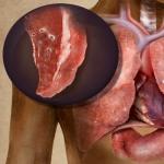


 Constellation Scorpio description")


